X-ray fluoroscopic system EXTRON 7, (C-Arm) produced by DRTECH Corp. (Republic of Korea)
X-ray fluoroscopic system EXTRON 7 manufactured by DRTECH (Republic of Korea) is intended for intraoperative X-ray control, minimally invasive and interventional interventions in various nosologies: in orthopedics and traumatology , abdominal surgery, urology and gynecology, vascular and cardiac surgery. The device can be used to work in operating rooms and trauma centers for continuous or periodic X-ray control.
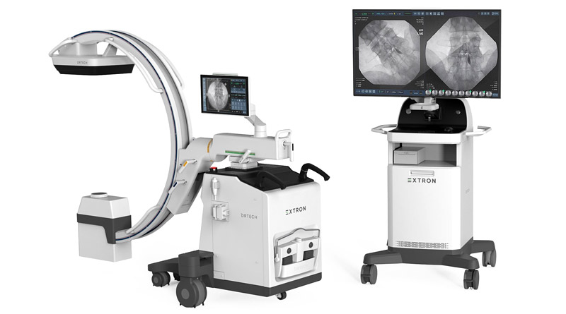
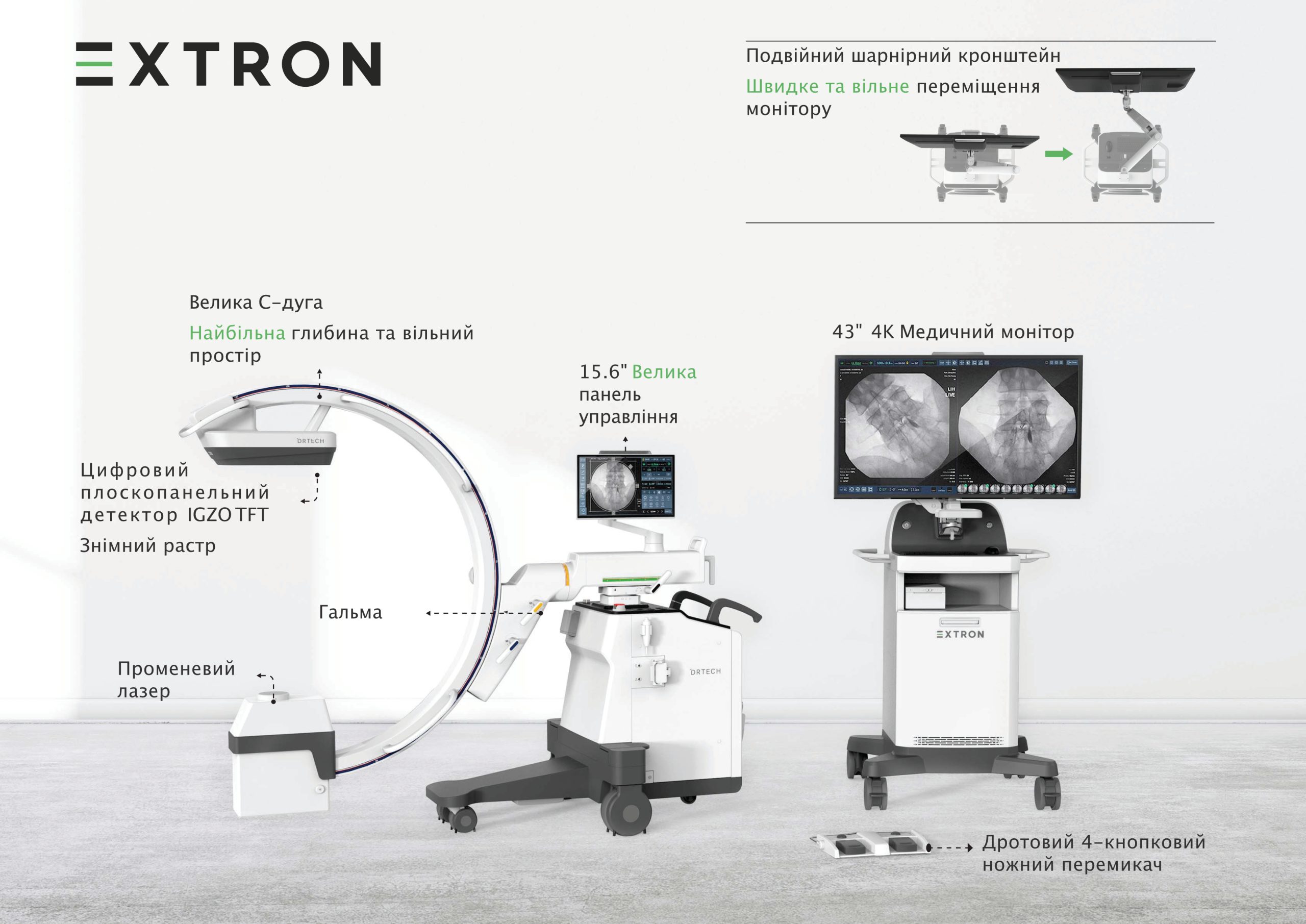
The EXTRON 7 surgical system consists of a mobile stand with a C-arm and an image processing workstation. The maneuverability of the system is ensured by strong wheels, and the brakes ensure the stability of the equipment and exclude accidental movements during its use or storage.
Any movement of the mobile stand’s C-arm (from rotation to orbital movement) can be locked using the lock switches.
Image quality of mobile X-ray system EXTRON 7
- The presence of a 30 x 30 cm digital detector makes it possible to obtain a larger image compared to similar systems from other manufacturers.
- Provides high-resolution (148㎛) images [1×1 images at 25 frames per second (fps)].
Low Dose Technology
Moving function for perfect collimator control: diaphragm and 4 “petals” for precise collimation.
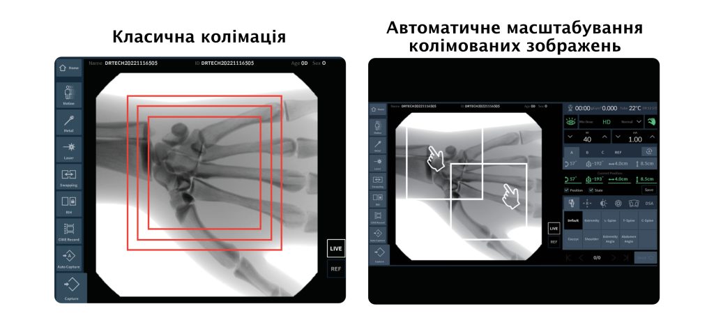
Automatic collimation screens only the initial state, while the EXTRON mobile x-ray system’s calibration movement function allows you to precisely control the collimator anywhere on the image for a perfect display.
Conventional C-arm fluoroscopic systems perform collimation using control buttons on the operator console, and the desired area can only be marked from the center of the square-shaped image.
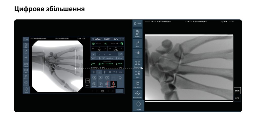
X-ray fluoroscopic machine EXTRON 7 can define imaging areas from any location and size by simply touching and zooming the desired image area on the control panel of the mobile C-arm stand. The rest of the area is collimated automatically to prevent excess exposure.
The selected image area is enlarged and automatically adjusted to the display size.
Image processing technology of the mobile X-ray fluoroscopic system EXTRON 7
RNR Technology (Real-time Noise Reduction) – DRTECH’s advanced recursive filtering is based on noise detection. While conventional systems apply a recursive filter to the entire image, resulting in image delay and noise, RNR detects object motion and applies a different recursive depth to each moving and stationary region. As a result, it reduces noise in specific locations in real time. And delivers exceptional image quality with less latency and noise on dynamic surgical imaging for perfect exposure.
 Auto brightness memory function for stable image brightness even if metal enters the image area, resulting in lower dose images.
X-ray generator/tube technology
- Boost mode for imaging large patients (up to 60 mA)
- Rotating anode tube to increase tube life
- Slim focus 0.3/0.6 for high resolution images with minimal image distortion
- Protection of the tube against water (IPX3)
Portable solution for ease of use
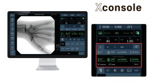
Direct work with digital images makes it possible to carry out accurate and detailed diagnostics using the tools of specially designed XConsole software developed by DRTECH .
Stored Positions feature stores position values for four axes simultaneously with dose information and image processing information to improve workflow efficiency and image accuracy. Also stores x-ray parameters for different C-arm positions and adjusts automatically, helping to reduce exposure to unnecessary dose and providing fast automatic exposure control (ABC Time).
Convenience for the user
- Stored positions for easy system positioning and dose control
- 4-button wired/wireless foot switch: 15 preset functions
- Large C-arm size: ∅1400 with a large depth for convenient patient and user movement
- 360° freely moving monitor
- Green laser for easy visualization
- Large control panel on mobile stand: 15.6“
DSA Angiography Package: (Vascular Studies)
Digital subtraction angiography (DSA) is a fluoroscopic research method used to clearly visualize the blood vessels of the cardiovascular system (vessels) in bone or soft tissues.
- Digital subtraction angiography (DSA)
- Roadmap (RSA)
- Re-mask
- Manual Pixel Shift
- Max. Peak turbidity (MSA)
- Landmark
- Application of special fluoroscopic and radiographic curves for individual modes of operation.
- Noise reduction, edge enhancement, auto cycle for all fluoroscopy modes, thumbnail view, contrast enhancement lookup table.
- Ability to store over 5000 digital images at maximum resolution in the DSA variant. Must be able to save images to CD/Pen drive etc.
- Edit frames for DSA images (video)
- Ability to export DSA images (videos) to mp4 format
Clinical areas of application of the EXTRON 7 fluoroscopic system
| Neurosurgery | Fusion (ALIF – Anterior Lumbar Interbody Fusion (Anterior Lumbar Interbody Fusion), TLIF, PLIF) PELD (endoscopic percutaneous lumbar laser discectomy) PECD (endoscopic percutaneous laser cervical discectomy) IDET (Intradisc Electrothermal Therapy) OLM (open lumbar microdiscectomy) ADR (cervical artificial disc replacement) ACF (cervical corpectomy with vertebral fusion) PVP (percutaneousvertebroplasty) |
|
Orthopaedics
|
CHS (compression hip screw) |
|
Anesthesiology
|
Epidural block |
|
Urology
|
URS (ureteroscopic stone removal)
Double-J ureteral stenting RGU (retrograde (ascending) uretography) RGP (retrograde (ascending) pyelography) Cystography
|
|
Gynecology
|
HSG (hysterosalpingography)
|
|
Diseases
internal
organs
|
EDP (retrograde cholangiopancreatography endoscopist) |
The device is equipped with a dosimeter, the use of which is mandatory, according to Article 17 of the Law of Ukraine “On the Protection of Humans from the Effects of Ionizing Radiation”.

